
Detailed discussions of the categories and subgroupings are given in Sections III,B through III,E. The scale of these drawings (Figs. 72-86) is approximately 20Å to the inch. β strands are shown as arrows with thickness, helices as spiral ribbons, and nonrepetitive structure as ropes. Disulfides are shown as "lightning bolts." Circles represent metals, and some prosthetic groups are shown as atomic skeletons, but not for all cases in which they are known to be present. A question mark in the label means that backbone connectivity is uncertain in some places. Where needed for clarity, the N-terminus of the domain is indicated by a small arrow; for a few two chain domains the C-terminus is indicated as well. [More detailed information on how the drawings were made is available in a Methods of Enzymology section (J.Richardson, 1985) and in a historical retrospective (J.Richardson, 2000 Nature Str. Biol.), and some related issues about structure representation in a review lecture (Richardson, 1992 Biophys. J.).]
[Protein structure schematics, figures 72 through 86, are presented in the web version as a gallery. That is, each figure is presented individually and navigation buttons to each figure and back to this page are available from each schematic. This allows a larger image for each schematic and faster loading in the browser. Links to each figure are given below:
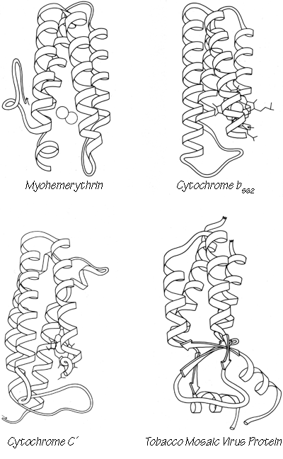
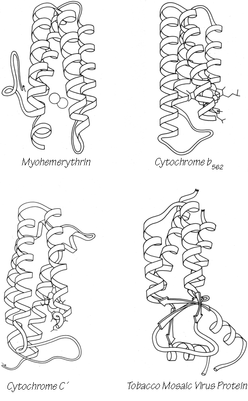
FIG. 72-1. Antiparallel α: up-and-down helix bundles.
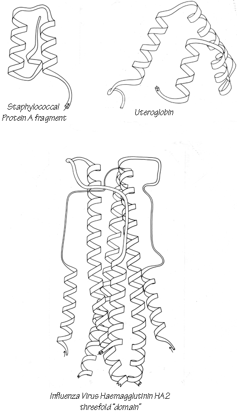
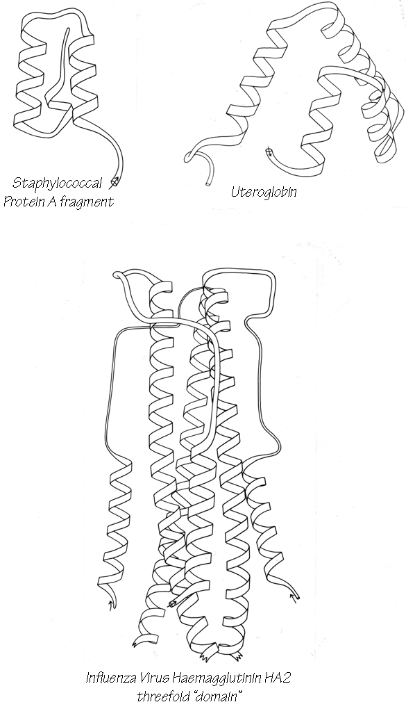
FIG. 72-2. Antiparallel α: up-and-down helix bundles.
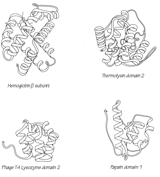
FIG. 73. Antiparallel α: Greek key helix bundles.
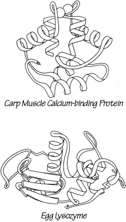

FIG. 74. Antiparallel α: miscellaneous.
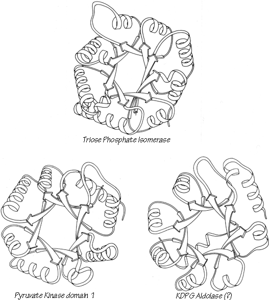
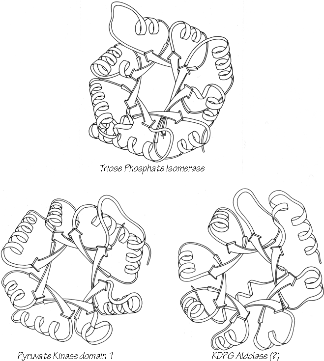
FIG. 75. Parallel α/β: singly wound parallel β barrels.
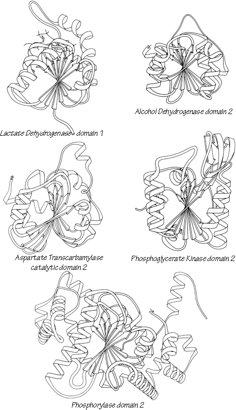

FIG. 76. Parallel α/β: classic doubly wound β sheets.
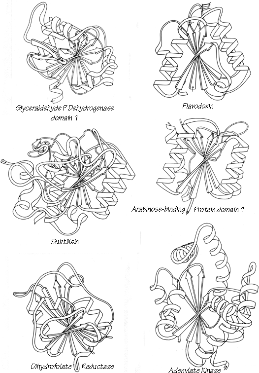
FIG. 77. Parallel α/β: doubly wound parallel β sheets.
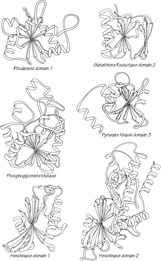
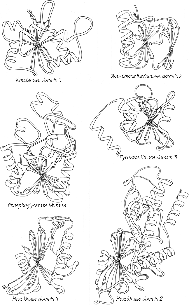
FIG. 77-2. Parallel α/β: doubly wound parallel β sheets.
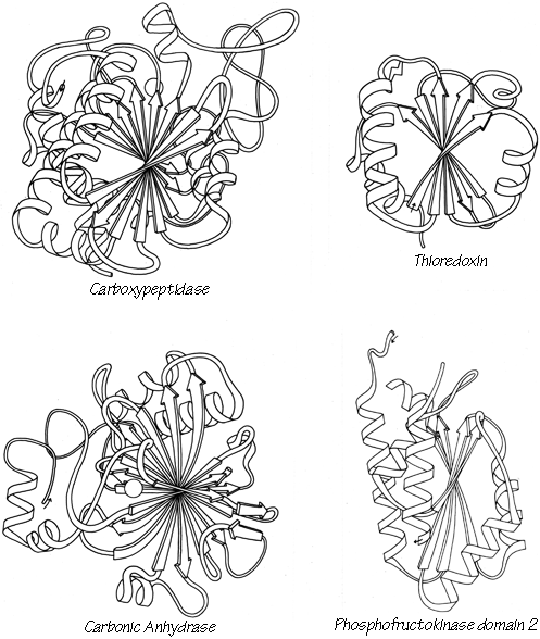
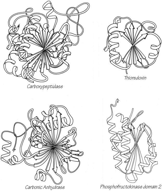
FIG. 78. Parallel α/β: miscellaneous.
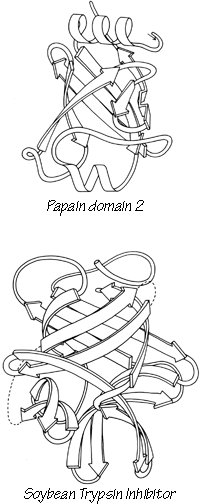
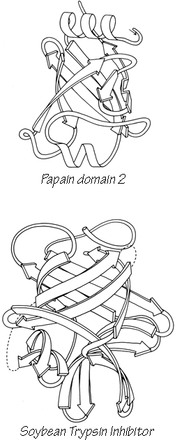
FIG. 79. Antiparallel β: up-and-down β barrels.
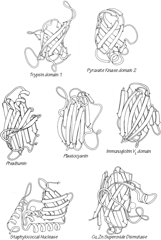
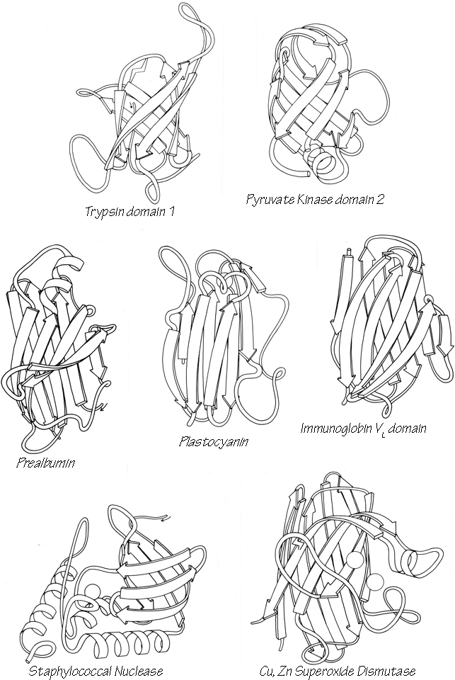
FIG. 80. Antiparallel β: Greek key β barrels.
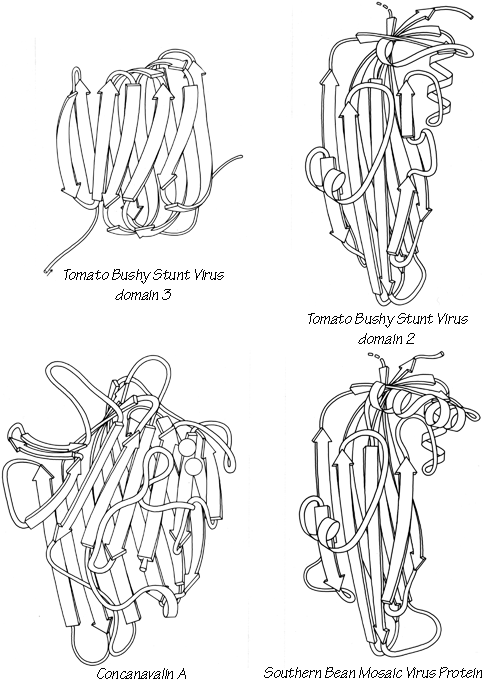
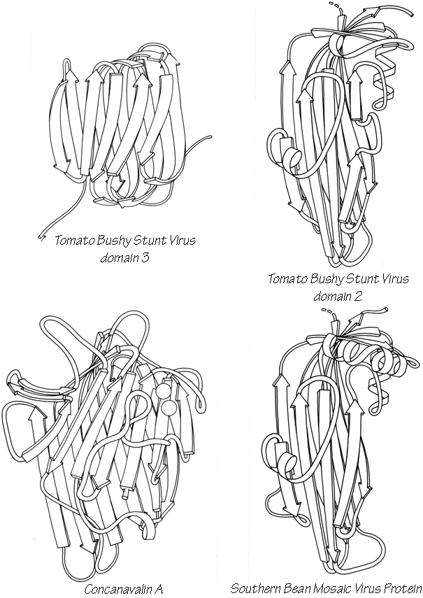
FIG. 81. Antiparallel β: "jellyroll" Greek key β barrels.
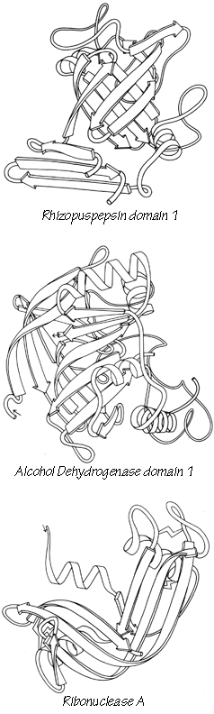

FIG. 82. Antiparallel β: other, multiple, and partial barrels.
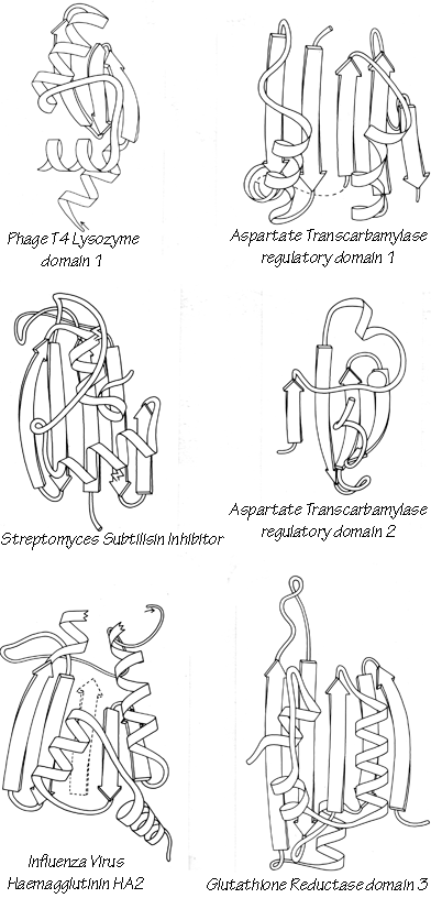
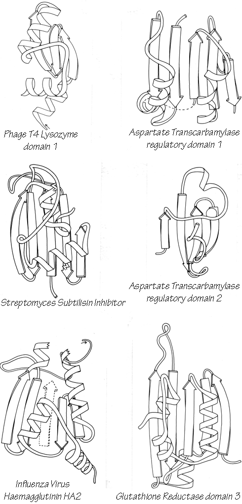
FIG. 83-1. Antiparallel β: open-face sandwich β sheets.
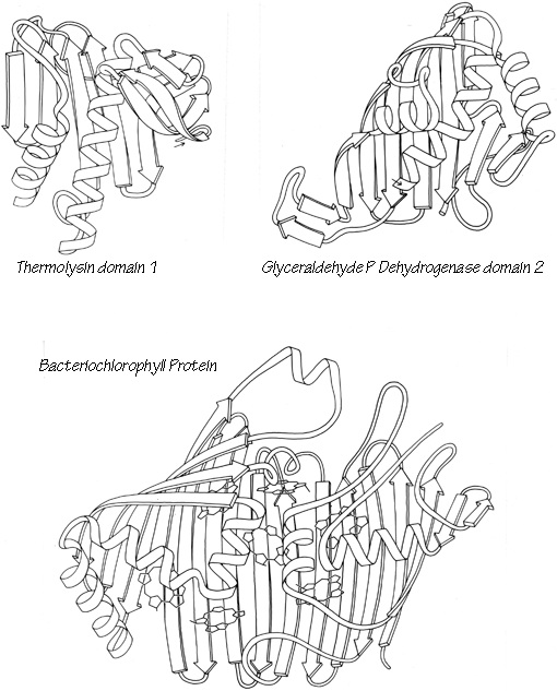
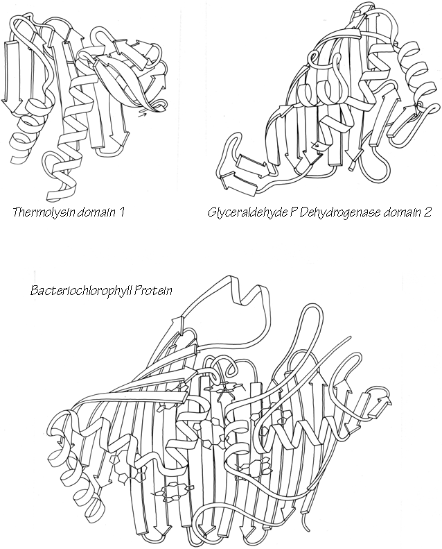
FIG. 83-2. Antiparallel β: open-face sandwich β sheets.
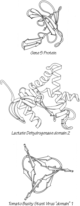
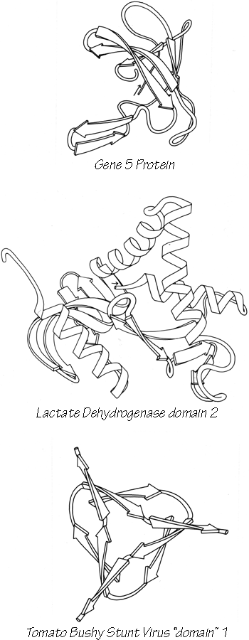
FIG. 84. Antiparallel β: miscellaneous.
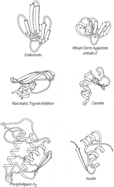
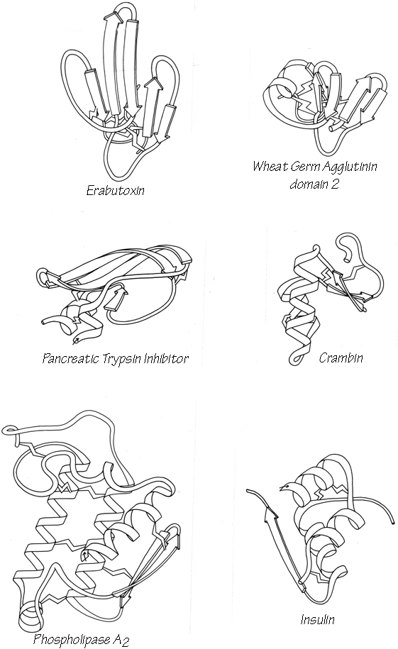
FIG. 85. Small disulfide-rich.

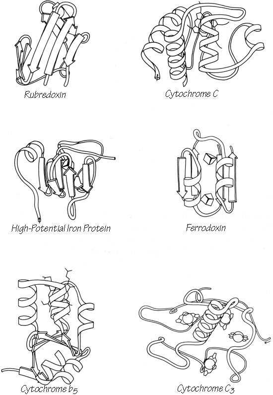
FIG. 86. Small metal-rich.